Anatomy Of Chest Area / Chest | Radiology Key / Ct anatomy of the chest, axial reconstruction.
byAdmin-
0
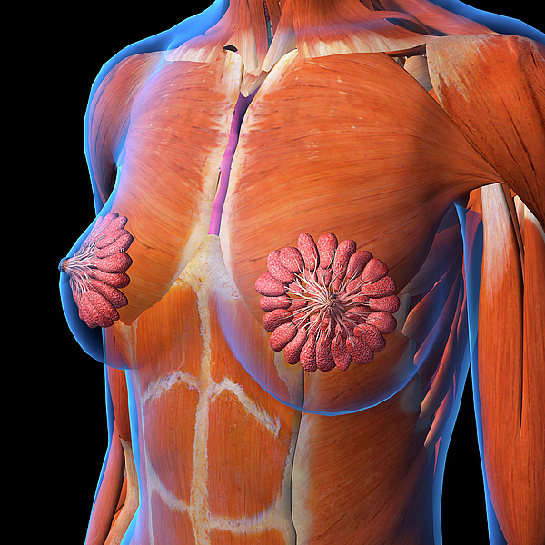
Anatomy Of Chest Area / Chest | Radiology Key / Ct anatomy of the chest, axial reconstruction.. Diagrams of normal venous anatomy of the thorax. Pathology of the heart, mediastinum, lungs and pleura. Manner of generating radiographic images, and technical. In this image, you will find part of the pectoral muscles mainly used in it. • a chest mri may be done for the following reasons:
Stability to arm and shoulder movement. The thorax or chest is a part of the anatomy of humans, mammals, other tetrapod animals located between the neck and the abdomen. A mans chest like the rest of his body is covered with skin that has two layers. A ) british informal used for referring to health problems in the area of your chest, especially … >> okay, so physical examination consists of four areas, inspection, palpation, percussion.
Radiological anatomy of chest including lungs,mediastinum ... from image.slidesharecdn.com You may also find triceps, lateral head brachialis anatomynote.com found chest muscle anatomy from plenty of anatomical pictures on the internet. Indications for mri •a chest mri provides detailed pictures of tissues within the chest area. Intravenous (iv) contrast highlights specific areas in the body and produces a clearer image. Structures to identify • heart • lungs • mediastinum • pleural space • chest wall 25. Meet your pectoralis major and pectoralis minor. Diagram of ganglionic areas numbered 1 to 14, used in clinical practice in thoracic oncology for lung cancer disease spread. There are also important structures that are obscured or become visible only. Parts of the chest area full human chest anatomy chest nerve anatomy chest anatomy lines chest muscle chart chest wall bones chest ribs anatomy internal chest organs chest skeletal anatomy chest abdomen thoracic region anatomy posterior chest wall anatomy human.
Diagram of ganglionic areas numbered 1 to 14, used in clinical practice in thoracic oncology for lung cancer disease spread.
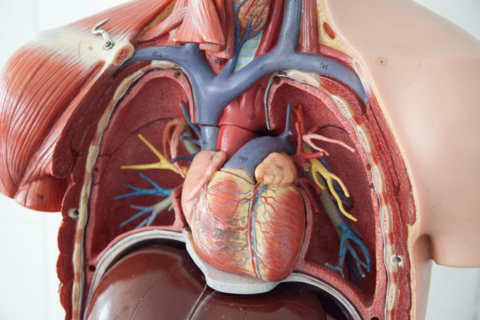
Structures that pass through this area can be thought of as the birds of the mediastinum: Radiology basics of chest ct anatomy with annotated coronal images and scrollable axial images to help medical students and junior doctors learning anatomy. This article is about the anatomical term. We think this is the most useful anatomy picture that. Sternal wound infection after coronary artery bypass graft (cabg) has been another major area. Learn about chest anatomy with free interactive flashcards. Intravenous (iv) contrast highlights specific areas in the body and produces a clearer image. Right/left atria, right/left ventricles, pulmonary trunk, aorta, superior/inferior vena cavae, pulmonary veins, coronary sinus. The hands should finish down low close to the hips to target this area of the pecs. 1, inferior lobe of right lung. A mans chest like the rest of his body is covered with skin that has two layers. Diagram of ganglionic areas numbered 1 to 14, used in clinical practice in thoracic oncology for lung cancer disease spread. The chest exercises are divided into barbell pressing exercises, dumbbell pressing exercises, machine pressing exercises.
• a chest mri may be done for the following reasons: Diagram of ganglionic areas numbered 1 to 14, used in clinical practice in thoracic oncology for lung cancer disease spread. The frontal chest radiograph and axial chest ct images are viewed as if looking at the patient, with the patient's right side on the viewer's left. Pathology of the heart, mediastinum, lungs and pleura. Chest muscles anatomy for bodybuilders.
Female Chest And Breast Anatomy Greeting Card for Sale by ... from images.fineartamerica.com Chest muscles anatomy for bodybuilders. Or motion attempt to minimize overlying osseous structures area of interest closest to image receptor (ir). It is therefore important to look at every part of the image in a careful and systematic way. A broad/hairy chest have you had any chest pains? Its anatomy is quite complex; 1, inferior lobe of right lung. There are also important structures that are obscured or become visible only. Chest — tʃest noun count *** 1.
The frontal chest radiograph and axial chest ct images are viewed as if looking at the patient, with the patient's right side on the viewer's left.
The hands should finish down low close to the hips to target this area of the pecs. Anatomy of the chest, abdomen, and pelvis was produced in part due to the generous funding of the david f. Swensen music we now show the physical exam of the heart. Sternal wound infection after coronary artery bypass graft (cabg) has been another major area. >> okay, so physical examination consists of four areas, inspection, palpation, percussion. The chest exercises are divided into barbell pressing exercises, dumbbell pressing exercises, machine pressing exercises. ■ identify the basic anatomy seen on a chest radiograph. Radiology basics of chest ct anatomy with annotated coronal images and scrollable axial images to help medical students and junior doctors learning anatomy. Anatomy of lung segmental anatomy of lung lateral view on a normal lateral view the contours of the heart are visible and the ivc is seen entering •a chest mri provides detailed pictures of tissues within the chest area. ) the upper front part of your body between your neck and your stomach: The major anatomical areas of interest on plain chest radiographs are however, abnormal radiographic appearances in the chest may be subtle and easy to miss. We think this is the most useful anatomy picture that. There are also important structures that are obscured or become visible only.
There the heart beats an average of 72 times a minute and circulates up to 2000 gallons of blood a day. Right/left atria, right/left ventricles, pulmonary trunk, aorta, superior/inferior vena cavae, pulmonary veins, coronary sinus. Its anatomy is quite complex; The major anatomical areas of interest on plain chest radiographs are however, abnormal radiographic appearances in the chest may be subtle and easy to miss. Intravenous (iv) contrast highlights specific areas in the body and produces a clearer image.
World's 1st procedure raises hopes for patients with leaky ... from medibulletin.com Iv contrast may be injected into a vein in the patient's arm or hand. Chest — tʃest noun count *** 1. Chest, abdomen, pelvisprovides detailed views of anatomic structures in general anatomy and function chest wall. We think this is the most useful anatomy picture that. Stability to arm and shoulder movement. Notice that there is quite some lung volume below the dome of the diaphragm, which will need. 1, inferior lobe of right lung. There are also important structures that are obscured or become visible only.
Diagrams of normal venous anatomy of the thorax. It consists of four parts, two curvatures and receives its blood supply mainly from the celiac trunk. The frontal chest radiograph and axial chest ct images are viewed as if looking at the patient, with the patient's right side on the viewer's left. 1, inferior lobe of right lung. Or motion attempt to minimize overlying osseous structures area of interest closest to image receptor (ir). Stability to arm and shoulder movement. Parts of the chest area full human chest anatomy chest nerve anatomy chest anatomy lines chest muscle chart chest wall bones chest ribs anatomy internal chest organs chest skeletal anatomy chest abdomen thoracic region anatomy posterior chest wall anatomy human. • a chest mri may be done for the following. Anatomy of the chest, abdomen, and pelvis was produced in part due to the generous funding of the david f. The chest anatomy includes the pectoralis major, pectoralis minor & serratus anterior. Chest muscles anatomy for bodybuilders. Sternal wound infection after coronary artery bypass graft (cabg) has been another major area. Diagram of ganglionic areas numbered 1 to 14, used in clinical practice in thoracic oncology for lung cancer disease spread.
Right/left atria, right/left ventricles, pulmonary trunk, aorta, superior/inferior vena cavae, pulmonary veins, coronary sinus anatomy of chest. Chest, abdomen, pelvisprovides detailed views of anatomic structures in general anatomy and function chest wall.